The Role of Ultrasound Imaging in Musculoskeletal Medicine
Non-invasive, real-time visualization of muscles, joints, tendons, and ligaments for precise diagnosis and treatment.
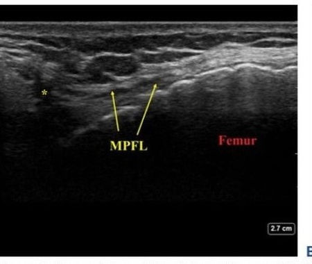
Ultrasound imaging has become a vital tool in the diagnosis and treatment of musculoskeletal disorders, contributing significantly to clinical practice in recent years. This non-invasive method uses a small probe, known as a transducer, to emit high-frequency sound waves that create spatial images of structures like muscles, tendons, joints, and ligaments. The transducer can be moved to capture different angles of the anatomy, providing live imaging, and still photographs when needed.
One of the key advantages of ultrasound over MRI is its ability to allow dynamic assessment, meaning the limb or joint being examined can be moved during the scan. This makes it easier to detect changes in the structure or function of muscles, tendons, and ligaments. Additionally, ultrasound guidance is essential in injection therapies, ensuring that treatments are delivered precisely to the correct location, optimizing outcomes.
In sports rehabilitation, ultrasound plays a crucial role in monitoring the healing process of injuries. Periodic imaging allows for tracking the recovery of lesions, enabling clinicians to adjust treatment plans based on real-time observations, leading to faster and more effective recovery. This makes ultrasound an indispensable tool for both diagnosing conditions and guiding therapeutic interventions.
Related Topics
Gallery


